A simple mockup that shows co-exploration of MRI data and CAD data. Controls are the same as before (press "W" and "S" to move the axial slice up and down):
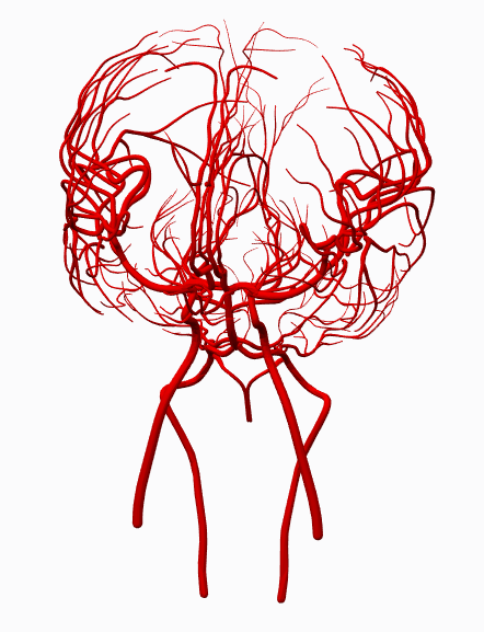
While assembling the basal ganglia CAD models, it occurred to me that I can also build a decent model of the arterial supply to the head. The arterial system was one of the first things we had to learn in the neurobiology course, so I'd like to assemble the 3D model.
Arterial system. Some details (e.g. posterior spinal arteries) are missing:
Superimposing the basal ganglia structures from yesterday:
There is an interesting website from U Tokyo, which hosts a repository of 3D models of various anatomical structures. I first found out about this website during a course on medical robotics, and am now encountering again in a basic neuroscience course. The site is called "BodyParts3D" and can be accessed here. Unfortunately, the site appears to be rather slow when accessing it from the US. Also, the navigation seems to be somewhat convoluted (at least for me). Try to figure out how to download specific 3D data!
I am currently interested in the anatomy of the deep nuclei in the human brain. Based on cross-sections presented in class, they appear to have an exquisite three-dimensional structure, and I'd like to get a better sense of the layout using 3D models.
In particular, I am interested in the following basal ganglia structures:
Caudate
Putamen
Globus pallidus
Subthalamic nucleus
Substantia nigra
Ventricular system
Internal capsule
For example, I was able to download the data for the left and right caudate nuclei (looks like something out of Alien):
A view of the ventricular system, with the "hole" in the middle presumably being space for the interthalamic adhesion:
Basal ganglia assembly (subthalamic nucleus, substantia nigra missing, because BodyParts3D doesn't have files for those structures). Also showing the middle cerebral artery:
Manipulating the CAD assembly:
Viewed from the bottom (gray structure in the midline is hypothalamus; gray structure directly medial to green / putamen is globus pallidus) The anterior, posterior limbs of the internal capsule is very visible, as is the genu (kink in the internal capsule). The thalamus (yellow) clearly abuts only the posterior limb of the internal capsule:
Transverse section:
Meanwhile, this is a nice diagram of the basal ganglia "circuit":
This term, I am taking a lab course on human neurobiology at Stanford. Among other things, we are learning about the anatomy of the human brain. I decided to take a look at my brain model / MRI data to see if I can identify some interesting structures.
First, I looked at the ventricular structures in the center of the brain. I do this by deleting the outer surface of the cortex in my previously generated 3D model. (Note that I may have to tweak the isosurface generation parameters from MRI data, in order to better segment internal structures.) The ventricular structure is roughly highlighted in green:
I think you can even find the foramina of Monro, which connects the lateral ventricles to the third ventricle:
Other things I am interested in are:
Do I have an intact Circle of Willis? (This will probably require resegmentation of the MRI model.)
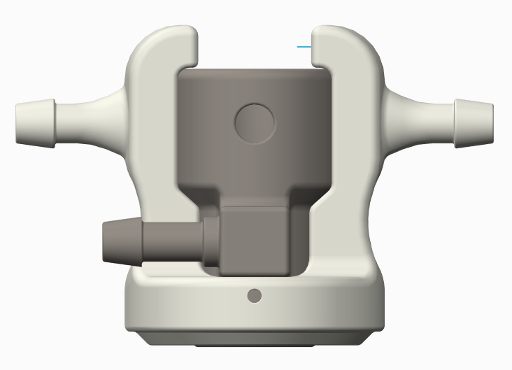
A part with "internal piping" in order to help recover leaked anesthesia from the Kopf small animal stereotax setup. The steel-colored part is a commercial Kopf part, and the white part surrounding it is my design: